|
> Home
> Research
> Publications
> Talks / Posters
|
As of February 2010, I am a postdoc at the University of Pennsylvania in Philadelphia, U.S.A., working on several problems in soft condensed matter theory and theoretical biophysics. With Andrea Liu and Philip Nelson, I study the development of early Drosophila embryos, in collaboration with the experimental group of Thomas Gregor at Princeton, and the development of early chicken hearts in collaboration with the experimental group of Dennis Discher at Penn. Together with Gareth Alexander I work on self-diffusiophoresis, the propulsuion of particles by gradients they themselves create. With Tobias Baumgart and Tom Lubensky I study the dynamics of phase separation in lipid bilayer membranes. Finally, I recently got interested in the collective behavior of self-propelled soft particles, in particular when they are close to jamming, which I study with the help of Carl Goodrich, Anshuman Pal and Isaac Carruthers.
Until December 2009, I worked at the Instituut Lorentz of Leiden University. There I worked with Cornelis Storm on two topics in biophysics: the morphology of membranes and the behavior of molecular motors. We were very lucky to be able to directly cooporate with the Leiden experimental biophysics group of Thomas Schmidt and the AMOLF/Leiden group of Marileen Dogterom on both subjects. At the same time, I also collaborated with the group of Jean-François Joanny at the Institut Curie in Paris on the topic of cell adhesion and contact inhibition.
In September 2012 I will move to a tenure track position at the new department of bionanoscience at the TU Delft.
|
Collaboration:
Julien Dubuis, Thomas Gregor
(experimental biophysics Princeton)
Lisa Manning, Andrea Liu, Philip Nelson
(theoretical biophysics UPenn)
|
|
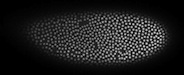
During the early stages of development of Drosophila embryos (the first 13 divisions), most nuclei populate the same cytosol without being divided into seperate cells by membranes. These nuclei are known as the syncytial nuclei. They populate the bulk of the embryo for cell divisions 1-8, after which most of them migrate to the surface, where we can visualize them by staining e.g. the histone in their DNA (see the site of the Gregor lab for movies). Using newly developed algorithms, we track the positions and shapes of the nuclei over time as they divide and form families of daughter nuclei. Studying the motion and shapes of the nuclei gives us a direct, non-penetrative handle on the dynamics inside the developing embryo, which allows us to study their mechanical properties in vivo. Moreover, we can combine these observations with simultaneous measurements of protein gradients, for example Bicoid, which is responsible for anterior-posterior symmetry breaking in the embryo. Doing so, we find emerging collective behavior, resulting from the local behavior at the level of the single nuclei.
|
Collaboration:
Gareth Alexander, Andrea Liu (UPenn)
|
|
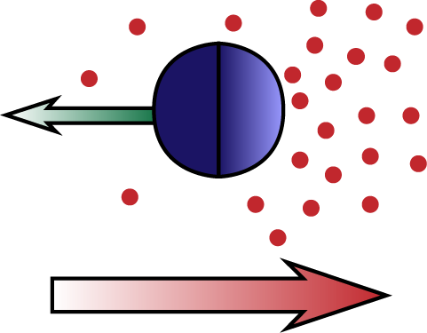
Diffusiophoresis is the process by which particles move trough a chemical concentration gradient, due to an attractive or repulsive interaction between the particle and the chemical compound. In self-diffusiophoresis the particle itself produces the compound it interacts with. An example is a particle that catalyzes the reaction 2H2O2 → 2H2O + O2, creating an O2 gradient and interacting with it. Another example is a particle that facilitates the polymerization of a biopolymer (e.g. actin), which creates a gradient because individual monomers diffuse, whereas the polymers do not. The latter process is one possible mechanism for the propulsion of Lysteria bacteria by means of actin 'comet tails'. We study these and related processes, analytically solving the Stokes equation for the fluid flow and extracting the particle motion in the diffusion dominated and advection dominated limits.
|
Collaboration:
Carl Goodrich, Anshuman Pal, Isaac Carruthers (UPenn)
|
|
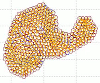
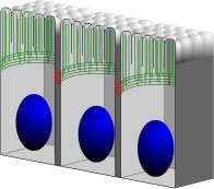
Soft particles are particles that can deform when they touch each other, resulting in a finite repelling force (rather than an infinite one, which is the case for hard particles). Both soft and hard particles undergo a jamming transition when their density gets above a certain critical value. When jammed, the particles have no degrees of freedom and thus can not move. We are interested in the case that the particles are self-propelled, which may both cause them to become jammed more quickly, and help them to break up a jammed cluster. To study this system, we use Brownian-dynamics like simulations as well as analytical techniques.
|
Collaboration:
Stefan Semrau, Thomas Schmidt
(experimental biophysics Leiden)
Cornelis Storm
(theoretical biophysics Leiden / Eindhoven)
References:
PRL 100, 088101 (2008)
Biophys. J. 96, 4906-4915 (2009)
PRL 104, 198102 (2010)
EPJE 34, 67 (2011)
|
|
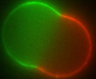
Membranes are everywhere in living cells - they form the boundary layer between the cell and its surroundings, and also the boundaries of the various organelles within the cell. The different membranes found in the cell exhibit a broad range of chemical compositions as well as a rich variety of (sometimes exotic) shapes. Over the last decade there has been a vivid discussion in the biophysics community as to whether the different molecules that make up the cell membrane (generically known as lipids) are organized into domains (called rafts) within the membrane or not. Such domains, if they exist in living cells, are too small to observe by optical microscopy; in artificial systems though, they can grow large and have indeed been seen.
Using differential geometry, we have developed a model for the shape of artificial membrane vesicles, which takes several properties of the membrane as parameters. Some of these can be measured directly, the remaining ones can now be obtained by fitting this model to the actual shapes, which we do in collaboration with the experimental biophysics group. The numbers we get out can be applied to living cells to determine conditions for domains to exist there and decide on which biological processes may be involved.
The large domains found in biomimetic vesicles also allow us to study curvature effects and membrane-mediated interactions between domains, comparable to the forces felt by proteins in living cells. These interactions can be quantified experimentally as well. We model them by again considering the effect the domains have on the curvature of the surrounding membrane. From this model we can explain both the measured interaction strengths and observed domain size distribution. Moreover, we find that the interaction has a striking effect: due to its nonlinearity, it spontaneously sorts membrane inclusions by size.
|
Collaboration:
J.M.J. van Leeuwen
(theoretical physics Leiden)
Reference:
Phys. Rev. E 80, 041924 (2009)
|
|
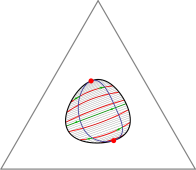
The Gibbs phase triangle (a section through the phase diagram at constant temperature and pressure) of a ternary lipid system exhibits a rich variety of phase coexistence regimes. Lipid mixtures can be in one of two liquid phases and a gel phase, and all of these can coexist, in pairs or even all three together. Especially intriguing is the recent discovery that there are systems for which each of the three underlying binary mixtures is uniform at a given temperature and pressure, but the ternary system exhibits phase separation. Extending Flory-Huggins theory for binary systems, we have developed a model that can explain all these different phase diagrams. Moreover, we can use our theory to predict the behavior of the line tension between coexisting phases as we move through composition space.
|
Collaboration:
Tobias Baumgart (experimental biophysics UPenn),
Tom Lubensky (theoretical physics UPenn)
|
|
Recent experiments in the groups of Tobias Baumgart (UPenn) and Patricia Bassereau (Institut Curie, Paris), have shown that pulling tubes from vesicles can induce nucleation of domains. For example, pulling a tube from the (stiffer) liquid ordered phase of a phase-separated vesicle initially produces a liquid ordered tube, but quickly nucleates a (softer) liquid disordered domain at the vesicle-tube neck. We study the dynamcis of this nucleation and phase separation process, both from a geometric and a thermodynamic point of view.
|
Collaboration:
Paige Shaklee, Thomas Schmidt, Marileen Dogterom
(experimental biophysics Leiden / AMOLF)
Cornelis Storm
(theoretical physics Leiden / Eindhoven)
References:
PNAS 105, 7993-7997 (2008)
Biophys. J. 99, 1835-1841 (2010)
|
|
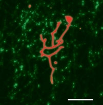
Molecular motors are the workers of the cell. They are the ones responsible for all transport processes, which can not rely on simple diffusion because it would be too slow. Molecular motors can 'walk' along the cell's 'roads' and 'highways' (the actin and microtubule networks) and increase distribution speeds to the necessary level. Moreover, motors also pull on the cell membrane and help create and maintain the non-equilibrium shapes of organelles such as the endoplasmic reticulum and the Golgi apparatus. Some motors, like Kinesin, are very efficient: a single one can take many steps along a microtubule before unbinding, and only a few are needed to transport a small vesicle from the nucleus to the cell perifery. However, there are also less efficient, so-called nonprocessive motors which unbind after taking only a single step. Amazingly, both types can pull membrane tubes, though the mechanism is quite different. Kinesin has been shown to dynamically cluster at the tip of a tube, where they cooporate to produce the necessary force to extract the tube from its mother vesicle. We have shown that, as part of this clustering process, kinesin motors get constantly recycled during tube pulling. Nonprocessive motors like Ncd on the other hand are a lot more dynamic. We found that these motors can create oscillating tubes, allowing for length regulation and exploration of different cell regions.
|
Collaboration:
Markus Basan, Martin Lenz, Thomas Risler, Jean-Francois Joanny
(Institut-Curie, Paris, France)
Reference:
Biophys. J. 98, 2770-2779 (2010)
|
|
When epithelial cells are placed on an elastic substrate and are given sufficient amounts of nutrients, they will grow along the substrate by extending filopodia. The force necessary for pushing the filopodia outward is provided by actin polymerization in the direction perpendicular to the cell's plasma membrane. During this growth stage the actin constantly grows and shrinks, and moreover branches and builds a network. When the cells have produced a confluent layer, the growth direction switches from parallel with to perpendicular to the membrane, until the cells mature at a height that depends on their specific type. The actin then no longer pushes on the membrane but instead forms a belt near the apical side of the cell. It reorients itself to be parallel to the membrane and forms bundles instead of branches. We investigate the process by which cells 'feel' the presence of a neighbor and switch from a growing state to a mature equilibrium state. Using reaction-diffusion systems, we study the role of several transmembrane and cytoplasmic proteins in this contact inhibition process, and also consider the possible ways in which it can go wrong, resulting in cancerous cell growth.
|
| |
|
|
> Liu group
> Nelson group
> UPenn physics
> UPenn
|
|