![]()
![]()
![]()
![]()
![]()
![]()
![]()
![]()
![]()
![]()
![]()
![]()
![]()

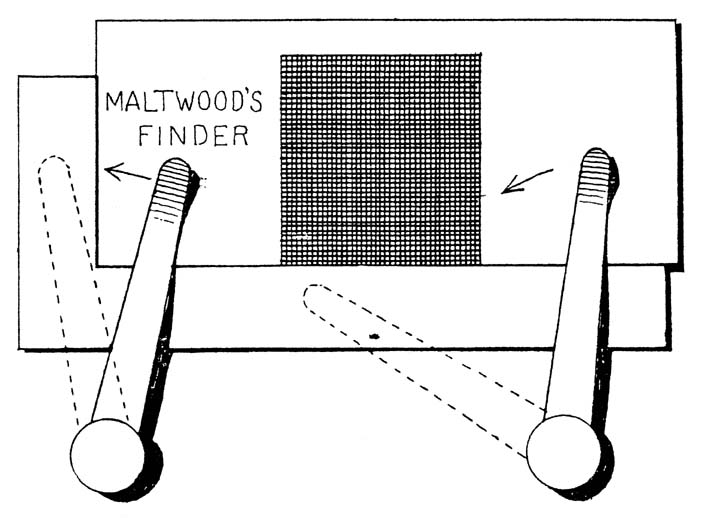
Substitute for the movable stage to be used with the maltwood finder, designed in 1908 by William Pepper, III, MD (M 1897). The device consists of a standard-size slide upon which a one-inch square grid is photographed. Two numbers code each of the grid's tiny squares. When the maltwood finder is placed over a blood or bacteriologic slide, the numbered grid system marks the location of a particular field or cell from the specimen, making it easy to return to an exact location on the slide for further investigation or photomicrographic reproduction. Pepper, grandson of the namesake for whom the William Pepper Laboratory was named, was an assistant professor of clinical pathology when he developed this device. For most of his career, he was a much-loved dean of Penn's School of Medicine. (JAMA, 20:2065, 1908)

Bacteria plate colony counter, as illustrated in Text-Book Upon the Pathogenic Bacteria (1896) by Joseph McFarland, MD (M 1889). Active as a bacteriologist within Penn's pathology department in the early 1890s and again from 1916 to 1936, McFarland authored one of the most used textbooks of its period. His Pathogenic Bacteria went through nine editions. (American Society for Microbiology Archives)
Koch's chamber for sterilizing and solidifying blood serum, illustrated in Abbott's Principles of Bacteriology (1892). (American Society for Microbiology Archives)


The rabbit test for rabies: plexiform ganglion of a rabbit dying of rabies produced by subdural inoculation. The capsules are filled or partially filled with foreign cells. In 1900 bacteriologist Mazÿck P. Ravenal, MD, and neuropathologist Daniel J. McCarthy, MD (M 1895), developed a more rapid way to diagnose rabies than the rabbit test, which required two to six weeks for results. Their method entailed examination of the intervertebral ganglia in the potentially rabid animal to detect the disease immediately. (University Medical Magazine, January 1901)
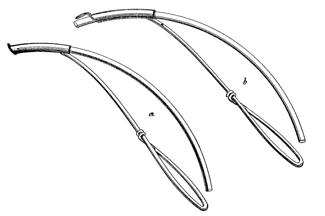
Curved cervical speculum for taking intra-uterine cultures: (a) tube with cap closed ready for insertion; (b) after insertion, showing the open cap and the projected inner tube for collecting the secretions. In 1908 Drs. William R. Nicholson (M 1893) and Joseph S. Evans (M 1899) developed this instrument in the William Pepper Laboratory and University Maternity Hospital to obtain clean specimens for examining bacteria in the uterus. Even then labs realized the difficulty and importance of obtaining proper specimens, still an issue with labs today. (Kelly's American Journal of the Medical Sciences, Vol. 136, 1908)
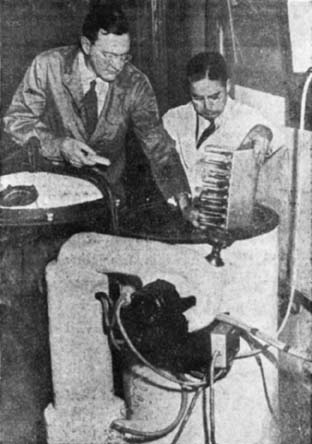
Drs. Stuart Mudd and Earl Flosdorf with their lyophilizer, a device for freeze drying blood plasma, circa 1935. Mudd and Flosdorf developed aseptic freeze drying techniques to preserve blood plasma, making it possible to give wounded soldiers on World War II battlefields blood transfusions through the use of dried plasma. Freeze drying, a practical invention for several industries, significantly impacted the clinical microbiology laboratory. The lyophilizer made it easier to preserve and disseminate bacterial strains for diagnostic and research purposes. Mudd began his Penn career on the Pathology faculty, then chaired the Department of Bacteriology (Microbiology) from 1931 to 1959. (University of Pennsylvania Archives)
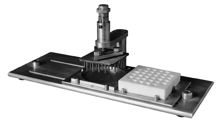
Steers replicator, invented in the William Pepper Laboratory by Drs. Edward Steers, Elwood L. Foltz and Betty S. Graves, 1959. This inoculum replicating apparatus for the routine testing of bacterial susceptibility to antibiotics advanced the study and detection of antibiotic resistance in human pathogens. A modified version of the device, found worldwide, remains in use in today's microbiology laboratories. In 1960 Steers headed up the Bacteriology (Microbiology) Section of the Pepper Laboratory.
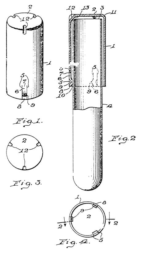
Morton's stainless steel culture tube closure, invented in the William Pepper Laboratory in 1939 and patented 1942. What appears to be a very simple, common device replaced the centuries-old laboratory standard - the cotton plug. (US Patent Office, #2,287,746)
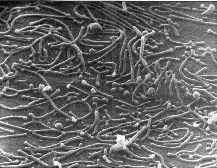
Scanning electron micrograph of Mycoplasma pneumoniae. In the early 1950s Drs. Harry E. Morton and Paul F. Smith collaborated on investigations of pleuropneumonialike organisms, now known as Mycoplasma pneumoniae, from the throats of humans. This work contributed to the large volume of literature Morton published on mycoplasms. (Phillips, D.M., N. Engl. J. Med. 331: 1421, 1994. ©1994 Massachusetts Medical Society)
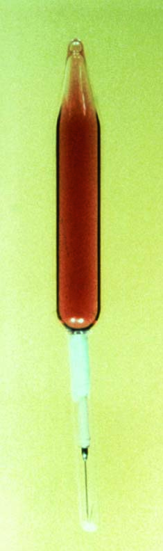
The first commercially available blood culture system, (Hynson, Westcott and Dunning, Inc., Baltimore), 1915. It utilized glass vacuum tubes manufactured by the Steele Glass Company of Philadelphia, and contained glucose bouillon with 1.5 percent sodium citrate as an anticoagulant. (CCW Judd and CE Simon, JAMA, 6 March 1915)
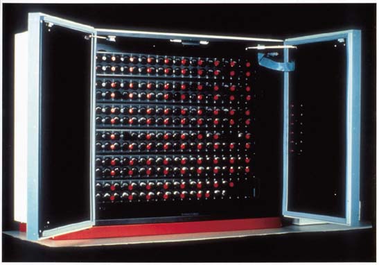
Modern day semiautomated blood culture instrument (left) used for continuous monitoring of microbial growth. Over 95 percent of positive blood cultures are detected with a 48- hour incubation period. (BD Bioscience#)
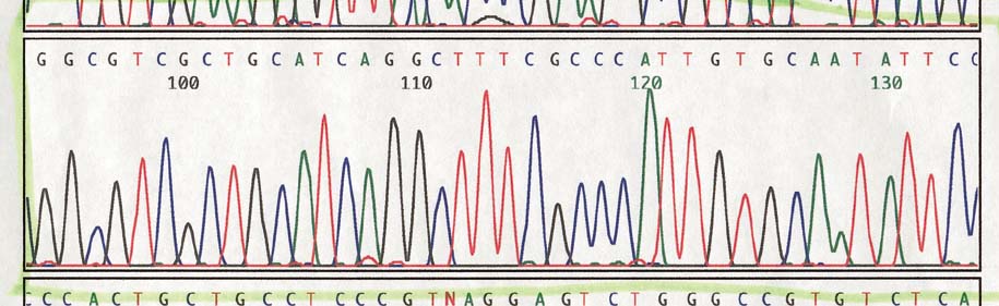
Four color sequence analysis of Mycobacterium sp. Rapid DNA sequencing, one of several advances in molecular biology and applications to diagnostic microbiology integrated into the William Pepper Laboratory in the 1990s, replaced lengthy biochemical tests. Polymerase chain reaction (PCR) technology also found a place in the lab for diagnosing many previously, difficult-to-diagnose infections such as TB and Herpes simplex encephalitis.