Color Vision
Under high light conditions, cone cells are responsible for much of our vision. Cone cells are very concentrated the fovea, but much less abundant in the retinal periphery.
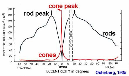
http://www.phys.ufl.edu/~avery/course/3400/vision/rod_cone_distribution2.jpg
Cone cells provide us with a number of visual advantages. First, their concentration in the fovea means that they are the cells responsible for providing us with fine visual detail. The fovea is the part of the retina that is receiving our direct visual attention. This region occurs over a very small angle in our visual field. The letters that you are reading right now are stimulating your fovea. However, if you do not move your gaze, the letter two lines up are not stimulating your fovea and are most likely impossible to read.
Cone cells are most unique in that they provide us with color vision. As mentioned ealier, there are three types of cone cells found in the retina, L(red), M (green), and S (blue) cones. L and M cones are far more more abundant than S cones especially in the periphery.
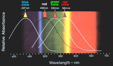
The diagram below shows the aborption curves for each of the photopigments in both cones and rods. Notice that there is some overlap in cone activation. This is important.
http://mcdb.colorado.edu/courses/3280/lectures/class14-2.html
Now, suppose an incoming light signal has a wavelength of 580 nm. This will cause both red and green cones to become activated while blue cones are not activated. Notice that this is the only point on the graph where these two relative levels of activation occur (see below). This is what gives rise to color vision. At almost every point on the visual spectrum, at least two different cones are stimulated. Their relative stimulation is what allows us to differentiate the wavelength of visible light based on color. In the areas where there is only one photoreceptor receiving light, it is impossible to differentiate color.
 Some individuals are born without the ability to make either red or green photopigments. This condition is more common in males than females because the gene for each of these photopigments resides on the X chromosome. Males only have one X chromosome while females have two. If a males lacks one of thees genes on his X chromosome he cannot make the red or green photopigment. On the other hand if females lack one of these genes on one X chromosome, it may be present on the other. Being unable to make a photopigment has profound implications for color sensitivity.
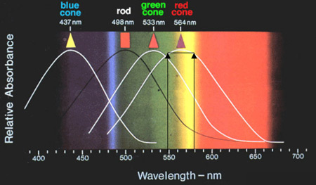
The relative activation of red and green cones by light at 580 nm
 If an individual lacking the M (green) cone is exposed to light at 580 nm, that light activates the red L (red) cone to the same extent as if was exposed to light at 550 nm in length (see left). This means the individual could not determine if the color they were viewing was orange or green. Individuals lacking L or M cones are said to be red-green colorblind because they have a difficult time distinguishing between red and green colors. To test to see if you may be red-green color blind click here.
The identical activation of the red cone by light of 550 and 580 nm.