Email me Jennifer Tareila Home
Project home
The Eye
Retinal And Rhodopsin
Absorption Spectra
WebMo Job 992
Applications
Links and Refs

The Eye And Vision


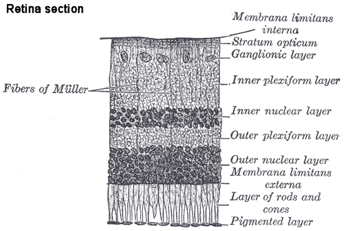
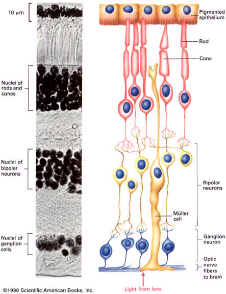
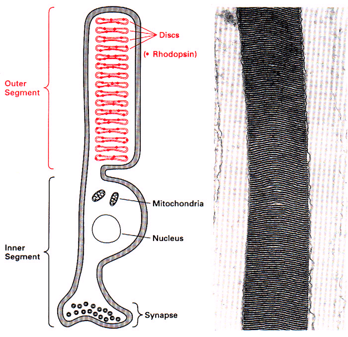
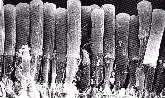
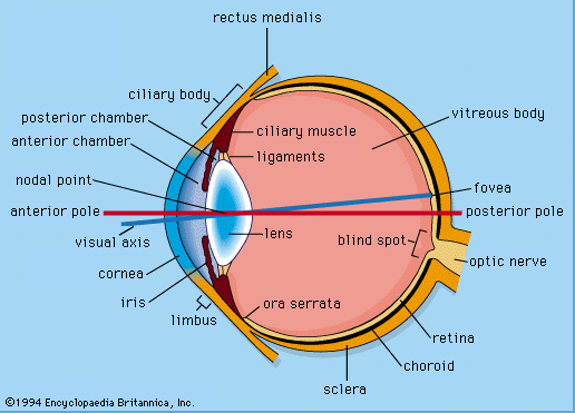
Figure from http://www.icat.ncsu.edu/projects/retina/files/eye.gif (ref page: http://www.icat.ncsu.edu/projects/retina/archive.htm)
   When light enters the eye, the photons first encounter the cornea, the covering of the eye.  The cornea will absorb any UV light, as it absorbs wavelengths below 315nm.  The remaining light passes through the cornea, and will be either reflected or absorbed 
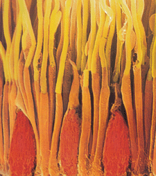
by the retina.  Light in the 400-700nm range is absorbed by either rods or cones, depending upon the specific wavelength involved.  Electromagnetic radiation longer 1000nm (infrared) is not absorbed- and a good thing it isn’t.  If we were able to see in this range, we would see heat, and almost everything would she shrouded in a fog of heat (15). 


   The human retina has approximately 100,000 rods and 3,000,000 cones.  The ratio of rods to cones is different in different species.  In mammals, the rods are approximately 1µm by 40µm. Rods have stacks of approximately 1,000 disks 16nm thick that contain chromophores. The inner segment contains the nucleus and typical cellular component, such as the mitochondria. The chromophore is contained in the outer segment of the rod. 
    Rods and cones absorb different wavelengths because the cis-retinal is joined to different proteins in these structures, so that the chromophore differs between them. There are 3 different proteins that the retinal can be joined to in the cones, which are responsible for color vision. Rods are responsible for vision in dim light.   Different animals have different proteins in their rods and cones.
    When light reaches the rods/cones and is absorbed, there is a conformational change made in the retinal molecule.  The molecule changes from the 11-cis-retinal isomer to the all-trans-isomer.  This change initiates a series of G-coupled protein changes that lead to a nerve impulse transmission, which results in vision. 
    The chromophores, or photo optically active portions, of the rod are composed of rhodopsin.    

Image retina section:(4)   http://cas.bellarmine.edu/tietjen/HumanBioogy/Sensory/sensor28.gif

Image (5) rods and cones in retina:  http://cas.bellarmine.edu/tietjen/HumanBioogy/Sensory/Image23.gif


Image(9) rod:  http://cas.bellarmine.edu/tietjen/HumanBioogy/Sensory/Image29.gif


Image (10)photomicrograph of rods: http://cas.bellarmine.edu/tietjen/HumanBioogy/Sensory/Image30.gif

scanEMphoto.jpe  from  http://webvision.med.utah.edu/photo1.html#pigments


Next to Retinal and Rhodopsin
Project home
The Eye
Retinal And Rhodopsin
Absorption Spectra
WebMo Job 992
Applications
Links and Refs