Email me Jennifer Tareila Home

Project home
The Eye
Back: Retinal And Rhodopsin
Next: Absorption Spectra
WebMo Job 992
Applications
Links and Refs

Retinal and Rhodopsin


http://www.theochem.uni-frankfurt.de/~birgit/Forschung/seite1.html
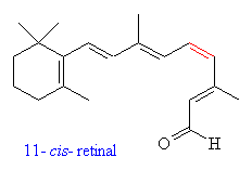
    Retinal is a polyene with four double bonds, and 16 possible isomers.  Only 11-cis-retinal is known to have any photoactivity in organisms.  Retinal was first noted in 1933 in photobleached bovine rhodopsin, which was observed to have a λmax of 385nm.  In 1944, the molecule was determined to be a vitamin A derivative. (25)
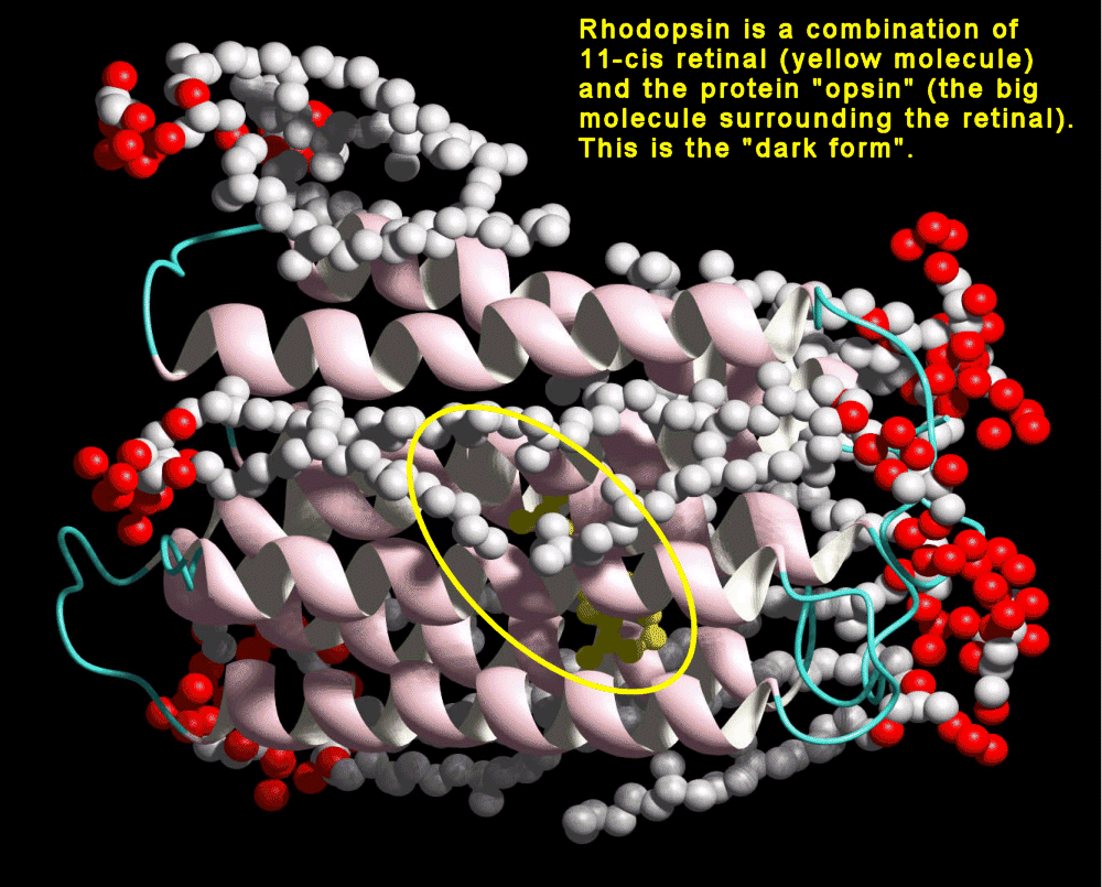
    In organisms, retinal is bound to an opsin protein. Rhodopsin, found in humans and other organisms, is a 40kD cross membrane protein with 7α helices and 348 amino acids. The carboxyl terminus resides in the cyctosolic side.  There are several different variants on the opsin molecules in organisms, and even slight differences in the cone cells of humans.


http://webvision.med.utah.edu/photo1.html#pigments
Rhodopsin, found in humans and other organisms, is a 40kD cross membrane protein with 7α helices and 348 amino acids. The carboxyl terminus resides in the cyctosolic side.  There are several different variants on the opsin molecules in organisms, and even slight differences in the cone cells of humans.


 http://www.science.siu.edu/microbiology/micr425/425Notes/09-Halobact.html
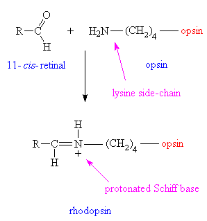
The retinal is bound to the opsin via a protonated Schiff base.


http://www.theochem.uni-frankfurt.de/~birgit/Forschung/seite1.html


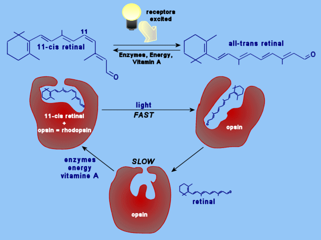
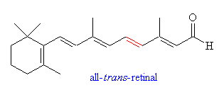
    When light brings about a photoreaction in the chromophore, the shape of the protein molecule is altered considerable, and Schiff base that connected the 11-cis-retinal to the protein is broken, leaving free all-trans-retinal in the chromophore.  At this point, the molecule is bleached; it has no absorption in the visible spectrum (25)  In response, the size is changed by about 5 Angstroms. (34). 
    The energy difference between the isomers is approximately 28KK (19, 29).  When the 11-cis-retinal is isomerized, the energy from the photon is converted into a motor action that then in turn initiates a cyclic GMP cascade that transmits a nerve impulse to the brain upon depolarization of the nerve cell membrane.  The different isomers have very different shapes, and these confomational changes are what initiates the cGMP cascade.  The cis isomer fits compfortably into the site (between the helices), while the trans isomer does not. The change starts the chain of events that leads to a nerve impulse.
    Different organisms have different opsins, and this is evident in the fact that many organisms see different wavelengths.  The differences in vision can be directly attributed to the differences in these opsins.  When the 11-cis-retinal is bound to an opsin, the delocalization of the electrons extends into the protein itself.     
    Rhodopsin in human rod cells is bound to scotopsin, an opsin protein.  Rhodopsin has a λmax of about 500nm.  (21, 34). 
Rhodopsins are easily excited by light as they are conjugated systems  that absorb in the visual range (28).


http://cas.bellarmine.edu/tietjen/HumanBioogy/Sensory/RhodopsinStructure.gif


Next to Absorption Spectrum
Project home
The Eye
Back: Retinal And Rhodopsin
Next: Absorption Spectra
WebMo Job 992
Applications
Links and Refs