


|
FLUORESCENT
WHITENING
AGENTS
IN LAUNDRY DETERGENT
& PAPER
|
| 1:
History 2:Importance and
Usage 3:
Spectroscopy 4: Current
Studies 5: Future Expts. |
| 6: HS Classroom
Lesson: A: Lesson
Plan B:
Worksheets C: Instructor
Guide |
 3.
Chemical Structure & Spectroscopy
3.
Chemical Structure & Spectroscopy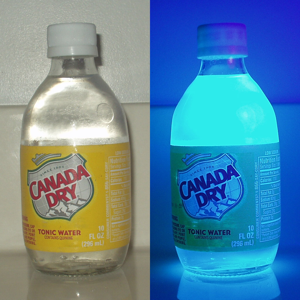 Those optical brighteners that are particularly
effective whitening agents, or FWAs, work in a manner similar to bluing
to
increase the perception of whiteness; they are colorless (do not absorb
visible light) and absorb
ultraviolet light, "converting" it to emit blue light
(1). Figure 1 to the left shows an example of a common
substance that exhibits these properties: quinine in tonic water.
Those optical brighteners that are particularly
effective whitening agents, or FWAs, work in a manner similar to bluing
to
increase the perception of whiteness; they are colorless (do not absorb
visible light) and absorb
ultraviolet light, "converting" it to emit blue light
(1). Figure 1 to the left shows an example of a common
substance that exhibits these properties: quinine in tonic water.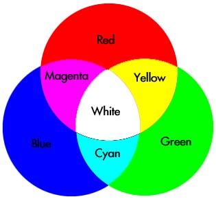 This emission of blue light "whitens"
fabrics that may naturally have a more neutral or yellowish white
tint. See Figure 2 to the right to recall how yellow and blue
light mix to create white light (remember this from Part 1?).
This emission of blue light "whitens"
fabrics that may naturally have a more neutral or yellowish white
tint. See Figure 2 to the right to recall how yellow and blue
light mix to create white light (remember this from Part 1?).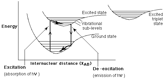 FWAs exhibit fluorescence as a result of
specific electronic and vibrational transitions, shown in Figure 3 to
the left:
FWAs exhibit fluorescence as a result of
specific electronic and vibrational transitions, shown in Figure 3 to
the left:
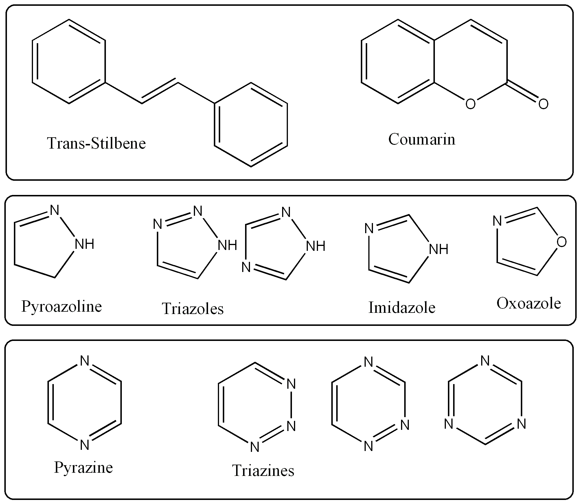 Most FWAs are
"derivatives of stilbene. . . biphenyl and five membered heterocyclics,
such as triazoles, oxoazoles or imidazoles. . . . six-membered
heterocyclics, such as coumarins, naphthalimide, pyrazine, or triazine
(7)." The
extensive pi-systems of these often heterocyclic aromatic compounds
are associated with the closely spaced electronic energy levels that
allow for energy transitions within the visible
range (e.g. n-->pi transitions). Table 1 to the right shows
some of base structures that
FWAs
are derived from.
Most FWAs are
"derivatives of stilbene. . . biphenyl and five membered heterocyclics,
such as triazoles, oxoazoles or imidazoles. . . . six-membered
heterocyclics, such as coumarins, naphthalimide, pyrazine, or triazine
(7)." The
extensive pi-systems of these often heterocyclic aromatic compounds
are associated with the closely spaced electronic energy levels that
allow for energy transitions within the visible
range (e.g. n-->pi transitions). Table 1 to the right shows
some of base structures that
FWAs
are derived from.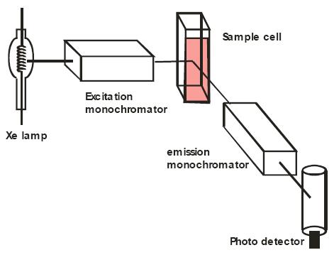 To
quantitatively characterize the absorption and
emission properties, it is most
To quantitatively characterize the absorption and
emission properties, it is most useful to use a fluorimeter to measure
the UV light absorption associated with
the first electronic transition and the visible light emission
associated with the second electronic transition. A fluorimeter
is
similar to a regular spectrometer except it must be outfitted so that
the light emitted by the sample cell must be carefulled filtered
(perhaps with slits or a diffraction grating) so that photodetector can
accurately assess absorption and emission of various wavelengths
(9).
Thus, fluorimeters are high-end, sensitive spectrometers. Figure
5 to the left shows how a fluorimeter operates and Figure 6 below shows
an actual fluorimeter.
To
quantitatively characterize the absorption and
emission properties, it is most
To quantitatively characterize the absorption and
emission properties, it is most useful to use a fluorimeter to measure
the UV light absorption associated with
the first electronic transition and the visible light emission
associated with the second electronic transition. A fluorimeter
is
similar to a regular spectrometer except it must be outfitted so that
the light emitted by the sample cell must be carefulled filtered
(perhaps with slits or a diffraction grating) so that photodetector can
accurately assess absorption and emission of various wavelengths
(9).
Thus, fluorimeters are high-end, sensitive spectrometers. Figure
5 to the left shows how a fluorimeter operates and Figure 6 below shows
an actual fluorimeter.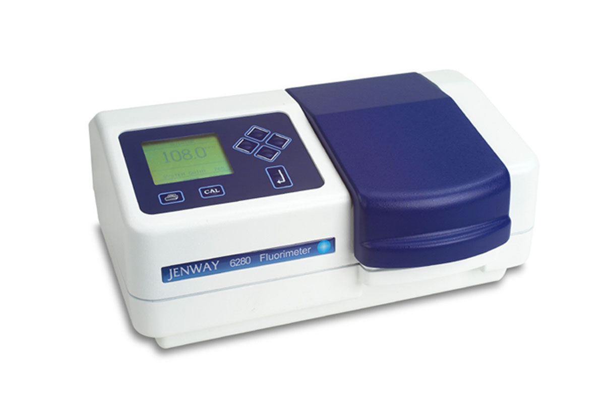 By calibrating the wavelength of the initial light
used for excitation
useful to use a fluorimeter to measure the UV light
absorption associated with
the first electronic transition and the visible light emission
associated with the second electronic transition. A fluorimeter
is similar to a regular spectrometer except it must be outfitted so
that the light emitted by the sample cell must be carefulled filtered
(perhaps with slits or a diffraction grating) so that photodetector can
accurately assess absorption and emission of various wavelengths
(9). Thus, fluorimeters are high-end, sensitive spectrometers.
By calibrating the wavelength of the initial light
used for excitation
useful to use a fluorimeter to measure the UV light
absorption associated with
the first electronic transition and the visible light emission
associated with the second electronic transition. A fluorimeter
is similar to a regular spectrometer except it must be outfitted so
that the light emitted by the sample cell must be carefulled filtered
(perhaps with slits or a diffraction grating) so that photodetector can
accurately assess absorption and emission of various wavelengths
(9). Thus, fluorimeters are high-end, sensitive spectrometers.|
Click here
to go to the index page for more spectra--the other
coumarins are not as well suited for use as FWAs because they absorb in
the
blue visible range, which would actually cause some yellowing, rather
than whitening and brightening. |

WORKS CITED:
|